
This video presents: 1. Cells taking part in the development of the conduction system of the hear. 2. Origin and development of the annulus fibrous (fibrous insulating ring). 3. Formation of the conduction pathway. 4. Difference of the His purkenje from the rest of the conduction system.
STUDY NOTES:
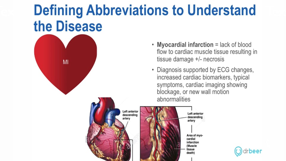
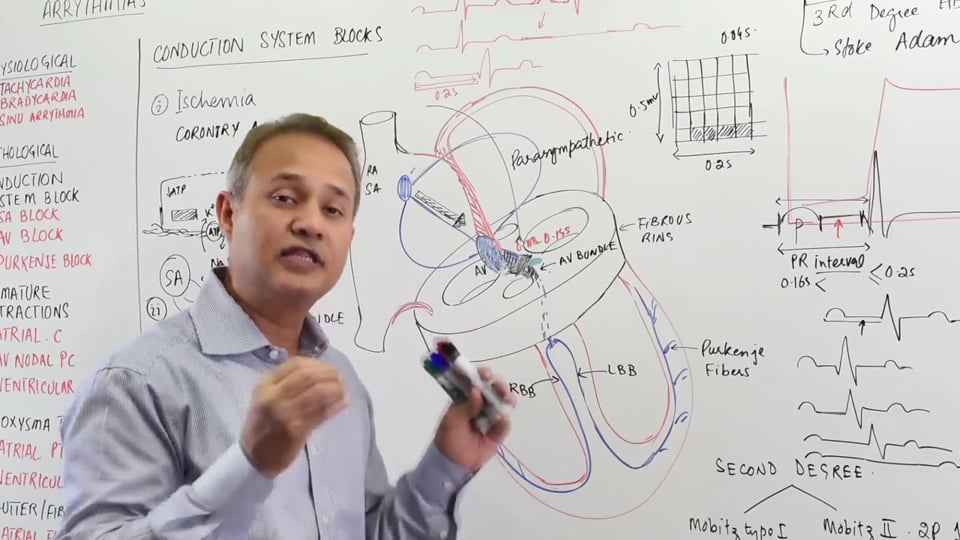
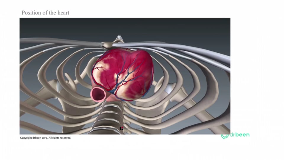
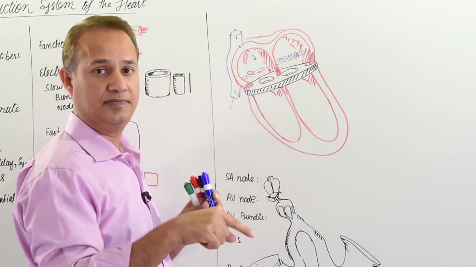
CONDUCTION SYSTEM OF THE HEART
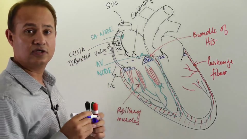
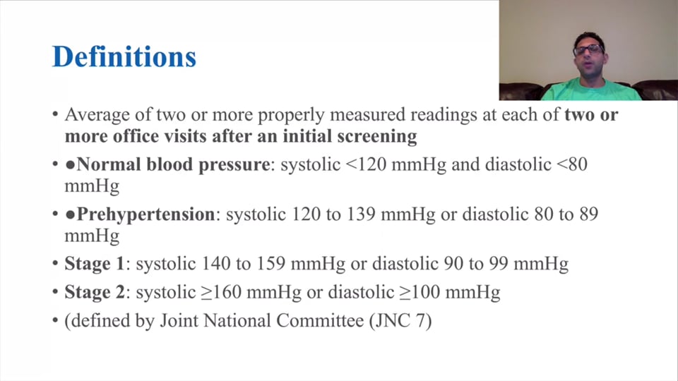
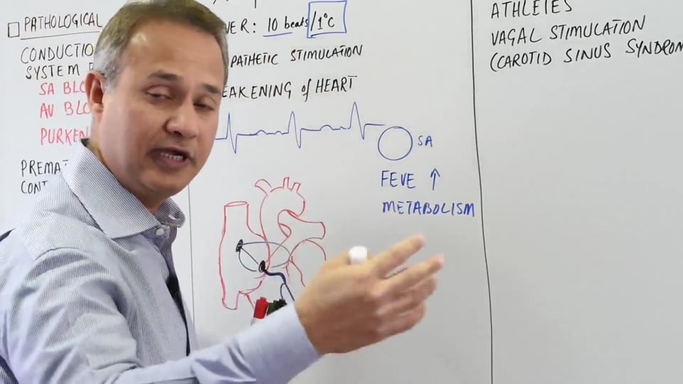
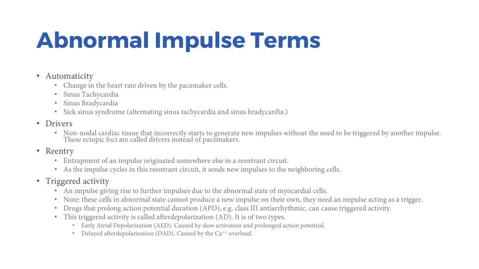
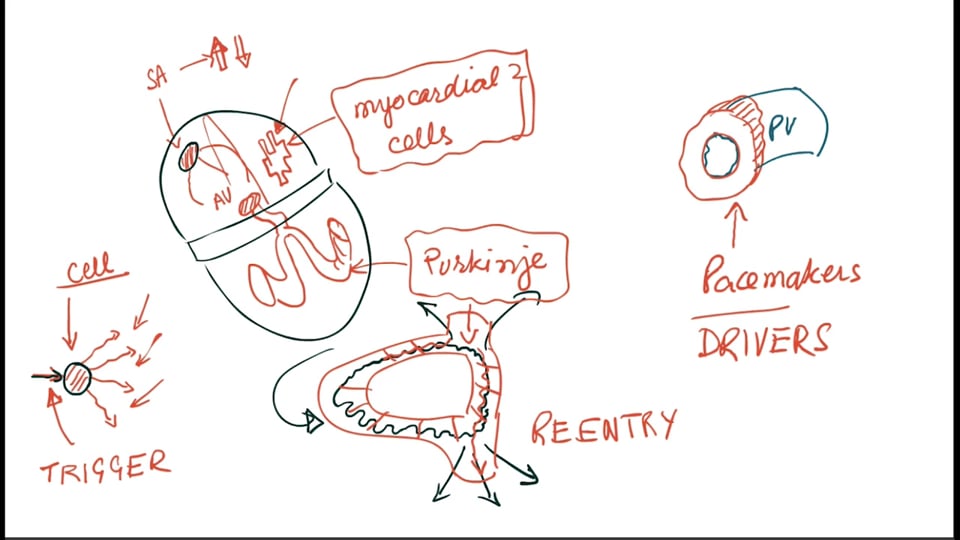
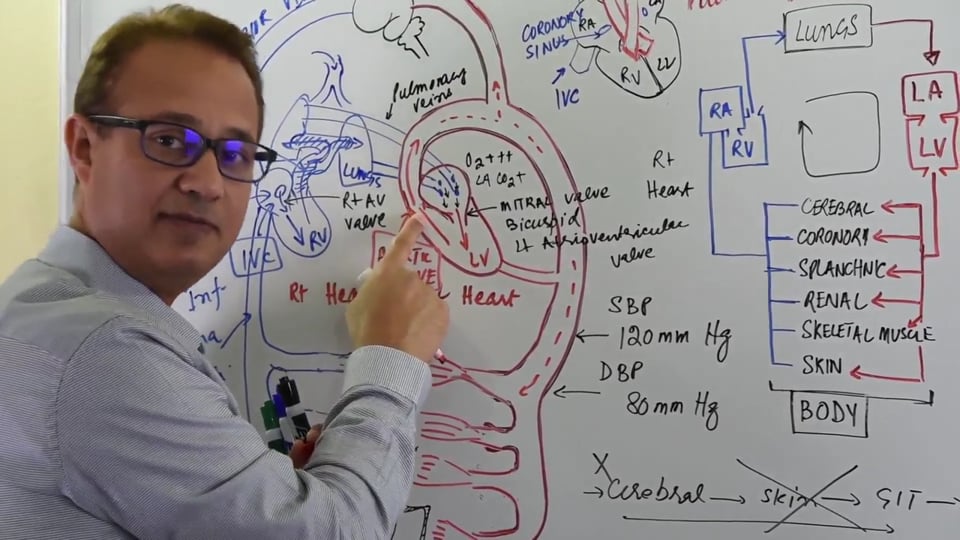
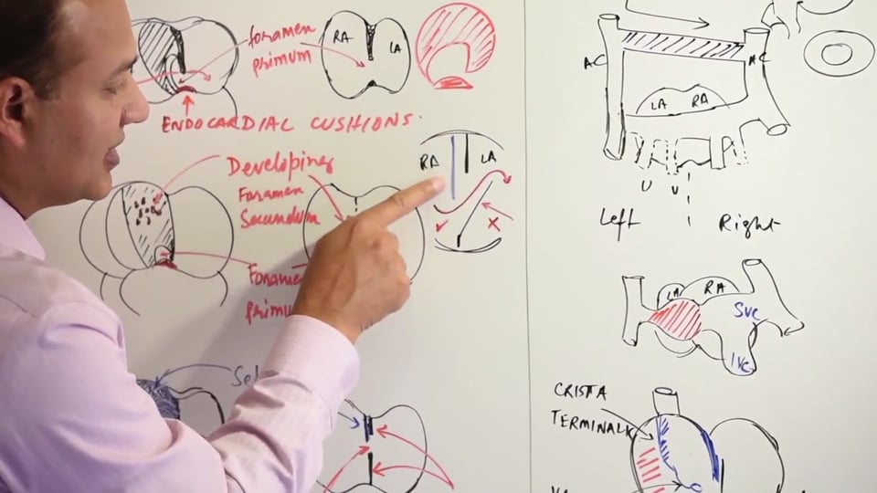
SAN which is present near the opening of the superior vena cava. SA nodal cells have the highest intrinsic rhythm of spontaneous depolarization (roughly 60- 100/min) which makes them the automatic choice for the pacemaker of the heart.
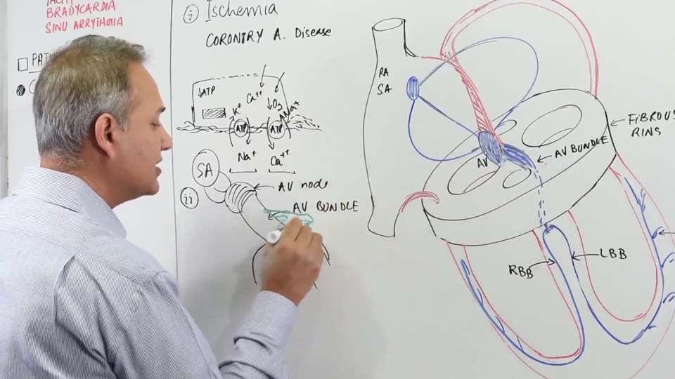
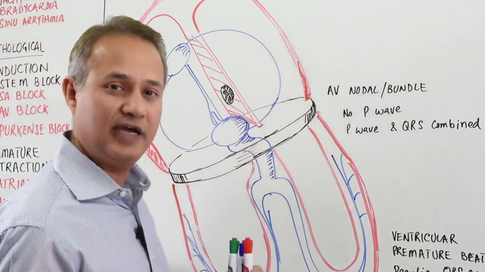
The AVN is present behind the endocardial cushions and in front of the coronary sinus. It's important to remember that the coronary sinus is actually the attritioned left horn of the sinus venosus. AV nodal cells have the second highest intrinsic rhythm (40-60/min). This automatically makes AVN the as the pacemaker of heart in case there's a damage to the SA nodal cells.
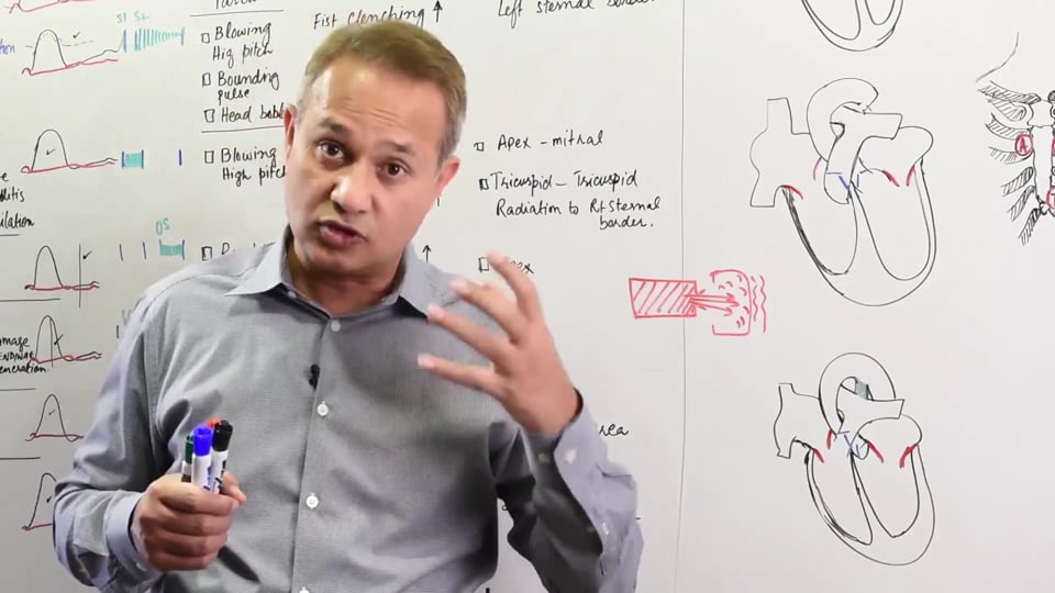
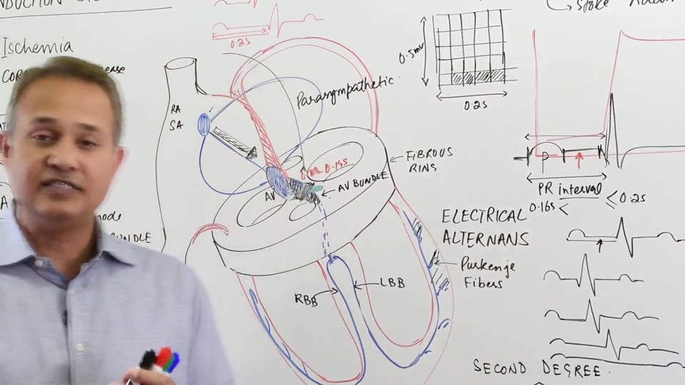
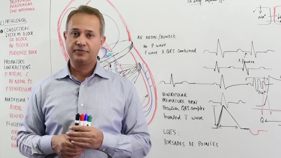
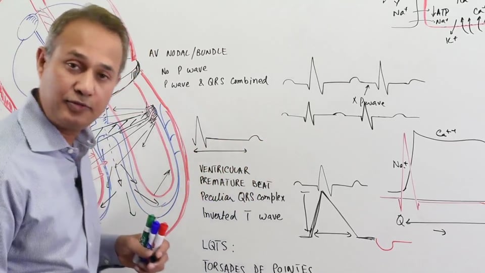
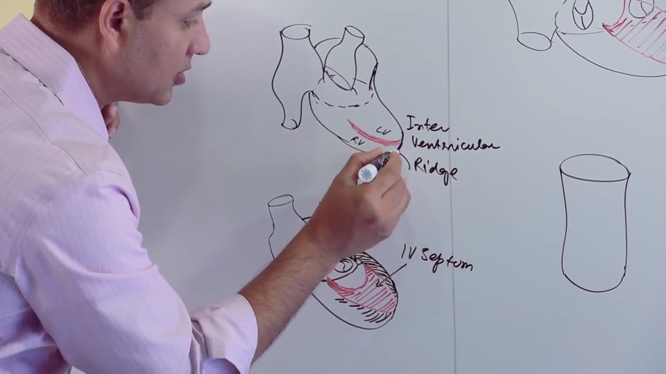
Bundle of HIS originates from the AV node and subsequently branches into two within interventricular septum. These two branches are the right and left bundle branches which ends up forming the HIS Purkinje system that supplies the papillary muscles and the rest of the ventricular myocardium. Papillary muscles are part of the trabeculated region of the ventricles which are derived from the primordial ventricle. Although, Purkinje cells are specialized for conduction only, they still possess an intrinsic rhythm of 35/min which gives them the property of automaticity. Hence, Purkinje system is third in line to take over as the pacemaker of the heart if anything goes wrong with both the SA and AV nodal cells.
The SA and the AV node develop from the sinus venosus. Before the sinus venosus gets incorporated into the right atrium and forms the conducting system of the heart, the primitive atrium serves as the function of the pacemaker.Atrial myocytes around the sinus venosus develop a faster intrinsic rhythm thereby naturally taking over as the pacemaker cells. These myocytes are derived from mesoderm.This means that as the myocardial cells are developing to form atria, they develop this ability to depolarize spontaneously. This allows the primitive heart to start beating by the 22nd day and that too without a true pacemaker, hence the primitive atria starts depolarizing even before the pacemaker is formed. Since sinus venosus is at the caudal end of the heart tube and serves as the inflow region. The initial pulsations are in coherence with the direction of the blood flow i.e., from caudal to the cranial side of the developing heart tube. Eventually as the sinus venosus is incorporated into the right atrium, the SA node develops from the sinus venosus near the entry of the superior vena cava.
The AV node also develops from the sinus venosus near the opening of the coronary sinus. As the AV node develops, bundle of HIS also develops along with it from the sinus venosus. The bundle of HIS develops within the interventricular septum and divides into right and left bundle branches. The cells around the AV node which become consolidated into forming the Bundle of HIS exhibit the MSX-2 homeobox gene. Purkinje fibers are actually modified contractile myocytes which start to function as conducting fibers when they become connected with Bundle of HIS cells.
Another important structure is the fibrous septum which insulates the ventricles from the depolarization of the atria and vice versa. This fibrous skeleton of the heart develops from the epicardium which is the visceral pericardium of the heart. The cells of the epicardium are derived from the local mesodermal cells around the sinus venosus as well.
This video presents the embryological development of :
1. SA node & AV node
2. Bundle of his
3. Purkinje system
Presented by Dr. Mobeen Syed
Following answers are created by ChatGPT. Occasionally the answer may be harmful, incorrect, false, misleading, incomplete, or limited in knowledge of world. Please contact your doctor for all healthcare decisions. Also, double check the answer provided by the AI below.
In addition to the presenter, following authors may have helped with the content writing, review, or approval:
ACCME Accreditation Statement
The DrBeen Corp is accredited by the Accreditation Council for Continuing Medical Education (ACCME) to
provide continuing medical education for physicians.
AMA Credit Designation Statement
The DrBeen Corp designates this enduring material for a maximum of 0.25 AMA PRA Category 1
Credits™.
Physicians should claim only the credit commensurate with the extent of their participation in the
activity.
In accordance with the disclosure policies of DrBeen Corp and the ACCME (Accreditation Council for
Continuing Medical Education), we are committed to upholding principles of balance, independence,
objectivity, and scientific rigor in all of our Continuing Medical Education (CME) and Continuing
Education (CE) activities. These policies include the careful management and mitigation of any relevant
financial relationships with organizations that are not eligible.
All members of the Activity Planning Committee and presenters have disclosed their relevant financial
relationships. The DrBeen Corp CE Committee has thoroughly reviewed these disclosures and determined
that these relationships are not deemed inappropriate in the context of their respective presentations.
Additionally, they are found to be consistent with the educational objectives and the integrity of the
activity.
| Faculty | Disclosures |
|---|---|
| Dr. Mobeen Syed | Author declares no conflict of interest. |
No credit card information needed.
 0.50 CME
Luis A Verduzco M.D.
 0.75 CME
Luis A Verduzco M.D.
Ahmed Zaafran, MD
 1.25 CME
Luis A Verduzco M.D.
Ahmed Zaafran, MD
 0.50 CME
Tatyana Travkina, MD
Ahmed Zaafran, MD
Ahmed Zaafran, MD
Ahmed Zaafran, MD
 0.75 CME
Tatyana Travkina, MD
Ahmed Zaafran, MD
Tatyana Travkina, MD
Ana Crawford M.D., M.Sc.
Ahmed Zaafran, MD
Ahmed Zaafran, MD
Ahmed Zaafran, MD
 1.25 CME
Dr. Mobeen Syed
Ahmed Zaafran, MD
 0.12 CME
Dr. Mobeen Syed
Ahmed Zaafran, MD
 1.25 CME
Dr. Mobeen Syed
Dr. Mobeen Syed
Dr. Mobeen Syed
 0.50 CME
Dr. Mobeen Syed
 0.75 CME
Dr. Mobeen Syed
Ahmed Zaafran, MD
 0.25 CME
Dr. Mobeen Syed
 0.16 CME
Dr. Mobeen Syed
 0.50 CME
Dr. Mobeen Syed
 0.16 CME
Dr. Mobeen Syed
Dr. Mobeen Syed
Dr. Mobeen Syed
 0.75 CME
Dr. Mobeen Syed
 0.50 CME
Dr. Mobeen Syed
 0.20 CME
Dr. Mobeen Syed
 0.20 CME
Dr. Mobeen Syed
 0.12 CME
Dr. Mobeen Syed
 0.09 CME
Dr. Mobeen Syed
 0.24 CME
Dr. Mobeen Syed
 0.25 CME
Dr. Mobeen Syed
 0.19 CME
Dr. Mobeen Syed
 0.08 CME
Dr. Mobeen Syed
 0.11 CME
Dr. Mobeen Syed
 0.09 CME
Dr. Mobeen Syed
 0.50 CME
Dr. Mobeen Syed
 1.00 CME
Dr. Mobeen Syed
 0.25 CME
Dr. Mobeen Syed
 0.50 CME
Dr. Mobeen Syed
 0.50 CME
Dr. Mobeen Syed
 0.50 CME
Dr. Mobeen Syed
 0.16 CME
Dr. Faraaz Bhatti
 0.50 CME
Dr. Mobeen Syed
 0.50 CME
Dr. Mobeen Syed
 0.50 CME
Dr. Mobeen Syed
 0.50 CME
Dr. Mobeen Syed
Dr. Mobeen Syed
 0.17 CME
Dr. Mobeen Syed
 0.50 CME
Dr. Mobeen Syed
Dr. Mobeen Syed
Dr. Mobeen Syed
Dr. Mobeen Syed
Dr. Mobeen Syed
Dr. Mobeen Syed
 0.50 CME
Dr. Mobeen Syed
Dr. Mobeen Syed
Dr. Mobeen Syed
Dr. Mobeen Syed
Dr. Mobeen Syed

Dr. Mobeen Syed

Dr. Mobeen Syed
 0.05 CME
Dr. Mobeen Syed
Dr. Mobeen Syed
 1.25 CME
Dr. Mobeen Syed
Dr. Mobeen Syed
Dr. Mobeen Syed
 0.50 CME
Dr. Mobeen Syed
 0.22 CME
Dr. Mobeen Syed
 0.50 CME
Dr. Mobeen Syed
 0.25 CME
Dr. Mobeen Syed
 0.13 CME
Dr. Mobeen Syed
 0.16 CME
Dr. Mobeen Syed
 0.16 CME
Dr. Mobeen Syed
 0.15 CME
Dr. Mobeen Syed
 0.15 CME
Dr. Mobeen Syed
 0.19 CME
Dr. Mobeen Syed
 0.75 CME
Dr. Mobeen Syed
 0.12 CME
Dr. Mobeen Syed
 0.16 CME
Dr. Mobeen Syed
 0.25 CME
Dr. Mobeen Syed
 0.24 CME
Dr. Mobeen Syed
 0.25 CME
Dr. Mobeen Syed
 0.19 CME
Dr. Mobeen Syed
 0.18 CME
Dr. Mobeen Syed
 0.50 CME
Dr. Mobeen Syed
 0.75 CME
Dr. Mobeen Syed
 0.50 CME
Dr. Mobeen Syed
 0.25 CME
Dr. Mobeen Syed
 0.50 CME
Dr. Mobeen Syed
 0.50 CME
Dr. Mobeen Syed
 0.25 CME
Dr. Mobeen Syed
 0.75 CME
Dr. Mobeen Syed
 0.13 CME
Dr. Mobeen Syed
 0.50 CME
Dr. Mobeen Syed
 0.25 CME
Dr. Mobeen Syed
 0.50 CME
Dr. Mobeen Syed
 0.50 CME
Dr. Mobeen Syed
 0.50 CME
Dr. Mobeen Syed
 0.25 CME
Dr. Mobeen Syed
 1.00 CME
Dr. Mobeen Syed
 0.25 CME
Dr. Mobeen Syed
0.20 CME
Dr. Mobeen Syed
 0.25 CME
Dr. Mobeen Syed
 0.17 CME
Dr. Mobeen Syed
 0.25 CME
Dr. Mobeen Syed
 0.50 CME
Dr. Mobeen Syed
 0.25 CME
Dr. Mobeen Syed
 0.19 CME
Dr. Mobeen Syed
 0.50 CME
Dr. Mobeen Syed
All information contained in and produced by DrBeen corp is provided for educational purposes only. This information should not be used for the diagnosis or treatment of any health problem or disease.
THIS INFORMATION IS NOT INTENDED TO REPLACE CLINICAL JUDGMENT OR GUIDE INDIVIDUAL PATIENT CARE IN ANY MANNER.
Click here for notice and disclaimer.
Write A New Comment
1 Comments
sanchezkenken964@*.com
Jan 21 2023, 5:32 pm
where do i start in what videos to learn EKG i am beginner level! please help or i will need to get refund